How can mini-organs help us to understand cancer?
Hear more from Maggie and Mimi about how growing mini-organs in the lab will help us to understand DNA signatures.
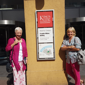
Maggie (left) and Mimi (right) at the entrance to the Franklin-Wilkins building, King’s College London
One of the great things about the Mutographs project is that we are using lots of different ways to explore the role of DNA signatures in cancer. One of the most exciting techniques being used involves growing mini-organs (also known as organoids) in a dish! The organoids work is led by Professor David Phillips and his team at King’s College London and is part of work area 4.
We visited the team at King’s to get answers to some of our questions about what organoids are and how they fit into the Mutographs project.
What causes DNA to change?
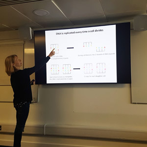
Dr Jill Kucab explains how DNA changes lead to cancer
After settling in with some coffee and generously sized muffins, we were treated to a series of talks that explained more about the work the team is doing.
Firstly Dr Jill Kucab explained about how chemicals can cause DNA to change. These changes leave distinctive patterns in DNA that are like a signature being left behind by the chemical. The Mutographs project aims to understand more about what causes these signatures.
Our cells need to split themselves in two in order to keep our bodies healthy. Most cells contain a copy of all of our DNA.
When the cell splits, DNA needs to copy itself so that both new cells have this complete set of DNA.
The cell has mechanisms to stop mistakes when it is copying DNA, but unfortunately they don’t always work. For example certain chemicals, like some of those in tobacco, can attach themselves to our DNA. This can cause the cell to copy the DNA wrongly and the new cell will end up with a mistake in its DNA. When the new cell splits, the mistake will be copied into more cells and so on. This can lead to cancer if the mistake happens in an important section of DNA.
What is an organoid?
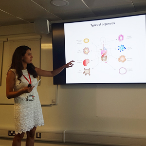
Dr Halh Al-Serori explains more about organoids
Next, we had an introduction to organoids from Dr Halh Al-Serori. Organoids are mini-organs that are grown in special dishes in the lab. The organs are grown from cells that people have donated.
What makes organoids so special is that they grow in three dimensions. This gives a researchers a good picture of how the cells might behave in the human body.
The organoids need to be kept in a special mixture of nutrients to keep them healthy, although it’s not always easy to guess what this mixture is! So far researchers have successfully created mini-organs of the pancreas, stomach, liver and lung. Other organoids, such as from the oesophagus, are proving harder to make as researchers still need to figure out the right formula of nutrients needed to keep them alive.
How are organoids being used in Mutographs?
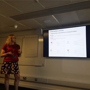
Dr Ellie Wilde explains more about work area 4 of Mutographs
Finally, we heard from Dr Ellie Wilde about what the King’s team have been doing so far. They are in the process of working out how the organoids respond to different chemicals linked to cancer. This will eventually help the team to understand how these chemicals change the DNA in the organoids, giving them a unique insight into how the DNA signatures are made.
The moon in a microscope
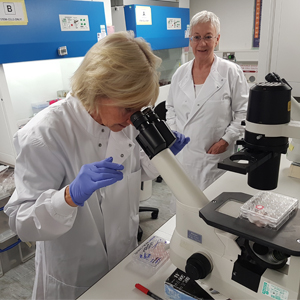
Looking down the microscope at mini-stomachs!
Once we’d heard about the project, it was on to the exciting bit – into the lab! Once we’d got our crisp white lab coats on, Halh showed us how they use a tank full of liquid nitrogen to store the cells used to make the organoids. This keeps them at a cool -196°C and means the cells can be kept for a long time.
We then went into the room where the cells are prepared for experiments. Ellie showed us how this is done under a special hood to stop the cells from being infected.
Then was the most exciting part, where we got to see the organoids themselves through the microscope! The ones that had been prepared were stomach organoids, which was timely because it was almost lunchtime. It was amazing to think that these tiny balls of cells scattered across the dish were tiny stomachs. It looked a lot like a moon inside the microscope!
With that, it was time to let the researchers get back to their important work. It was great to be able to understand more about organoids and see them up close. This area of work in the Mutographs project is essential to help teams across the project to understand more about how chemicals leave signatures in DNA. We are very much looking forward to hearing more from this great team as the project progresses.